 ‘The development of a novel MRI based method for measuring blood perfusion in neurovascular damage’ Emma Metcalfe-Smith
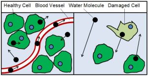
My work focuses on developing a new way to measure the movement of blood and water within body tissue using MRI (Magnetic Resonance Imaging). A greater understanding of this movement will help us to better understand the behaviour of different tissues. It is hoped that this will give more accurate measurements which will help in the diagnosis and treatment of injuries such as damage to nerves and blood vessels.
 ‘The Role of Diffusion Tensor Imaging in the Diagnosis of Paediatric Brain Tumours’ Heather Rose
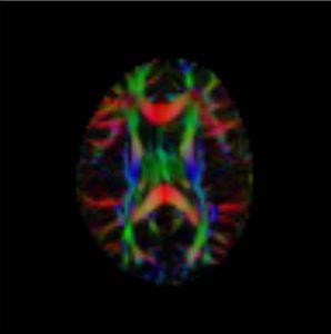
We focus on the use of advanced magnetic resonance imaging (MRI) to help diagnose brain tumours in young patients. We measure the movement of water molecules within the brain to help identify any change in structure in and around brain tumours. An additional focus of the research project is the design of databases that store the MRI images and allow researchers from different universities to work on joint research projects.
 ‘The Fusion of MRI and Diffusion Imaging (DTI) of the optic pathway as an aid to the management of Optic Pathway Glioma (OPG)’ Lara Worthington
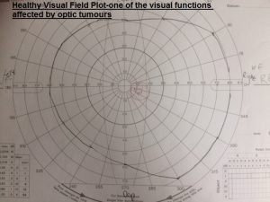
My research is aimed at trying to use MRI (Magnetic Resonance Imaging) to indicate possible damage to children’s vision arising from tumours located near the optical nerves in the brain.
‘Communicating information from MRI – Patient and parent views’ Natalie Tyldesley-Marshall
I am interviewing young patients and their parents about how they feel about, understand and value seeing the MRI (Magnetic Resonance Imaging) images of their / child’s brain tumour. We hope this will help better guide discussions between doctors and patients around their test results.
 ‘Measuring blood flow in childhood brain tumours using magnetic resonance imaging’ Stephanie Withey
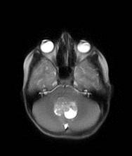
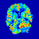
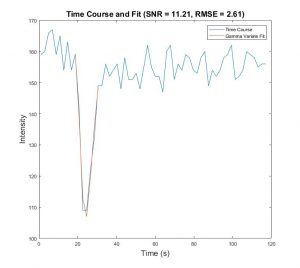
My research involves measuring blood flow in childhood brain tumours using magnetic resonance imaging (MRI). While patients are having their scan they are given an injection of a dye which enters the brain tumour and changes how it looks on the pictures from the MRI. I write computer programs that use these changes to give us extra information on the type of tumour it is and how well the treatment is working.
 ‘Development of novel methods for obtaining robust biomarkers from diseased and ageing brains’ Stephen Powell
My PhD focuses on developing methods to automatically assess image quality and obtain biomarkers from MRI (Magnetic Resonance Imaging). Biomarkers are quantities which can be measured and used to determine whether the brain is healthy or not.
 ‘The cutting-edge research of the advanced MR imaging technology in biomedical and brain science’ Yu Sun
My research focuses on using MRI (Magnetic Resonance Imaging) to better understand tumours in children and how to identify these earlier. My project looks at how to use the latest A.I. (Artificial Intelligence) to investigate brain disease and improve chances of survival.